Click on each image for a larger pop-up and
check-out the relevant details in the publication linked on the right of the picture.
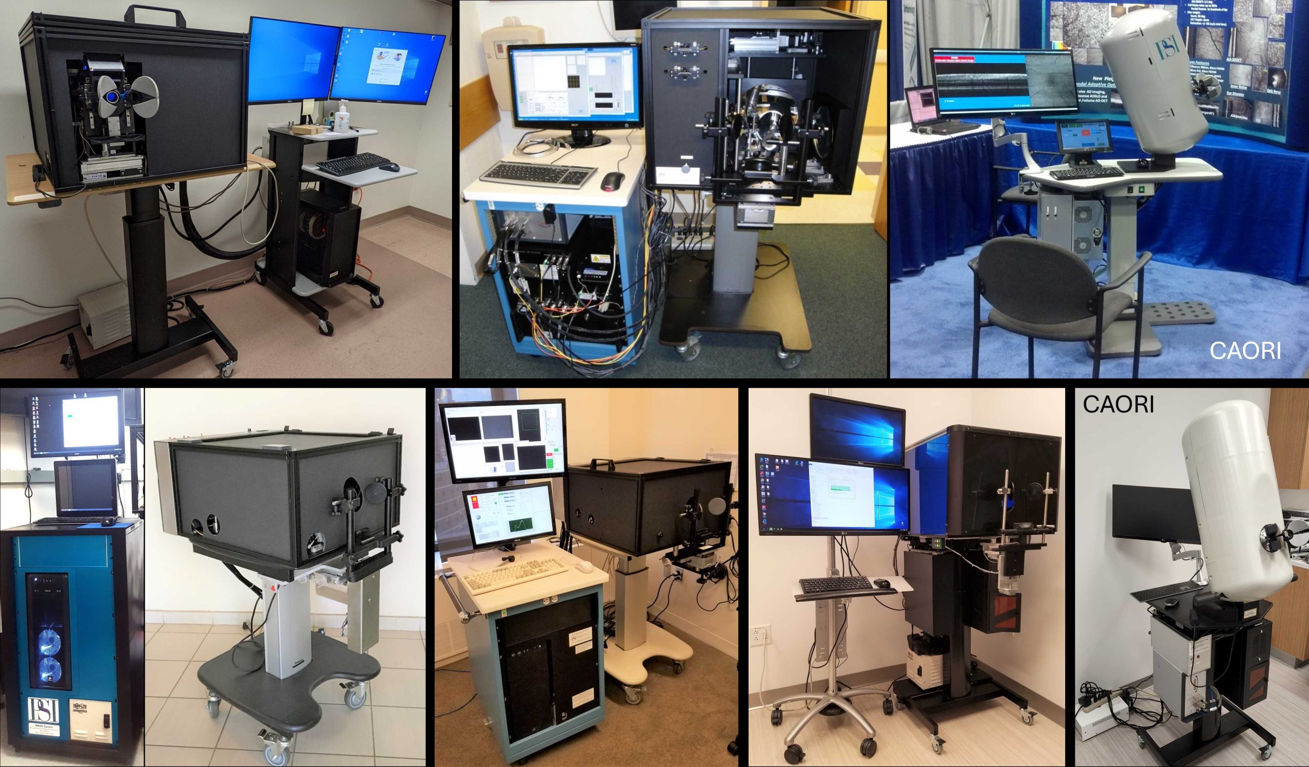
Examples of multimodal imaging platforms developed at PSI for human and small animal retinal imaging. | “High-Resolution Retinal Imaging: Technology Overview and Applications”, Photonics 11(6), 522, (2024) | |
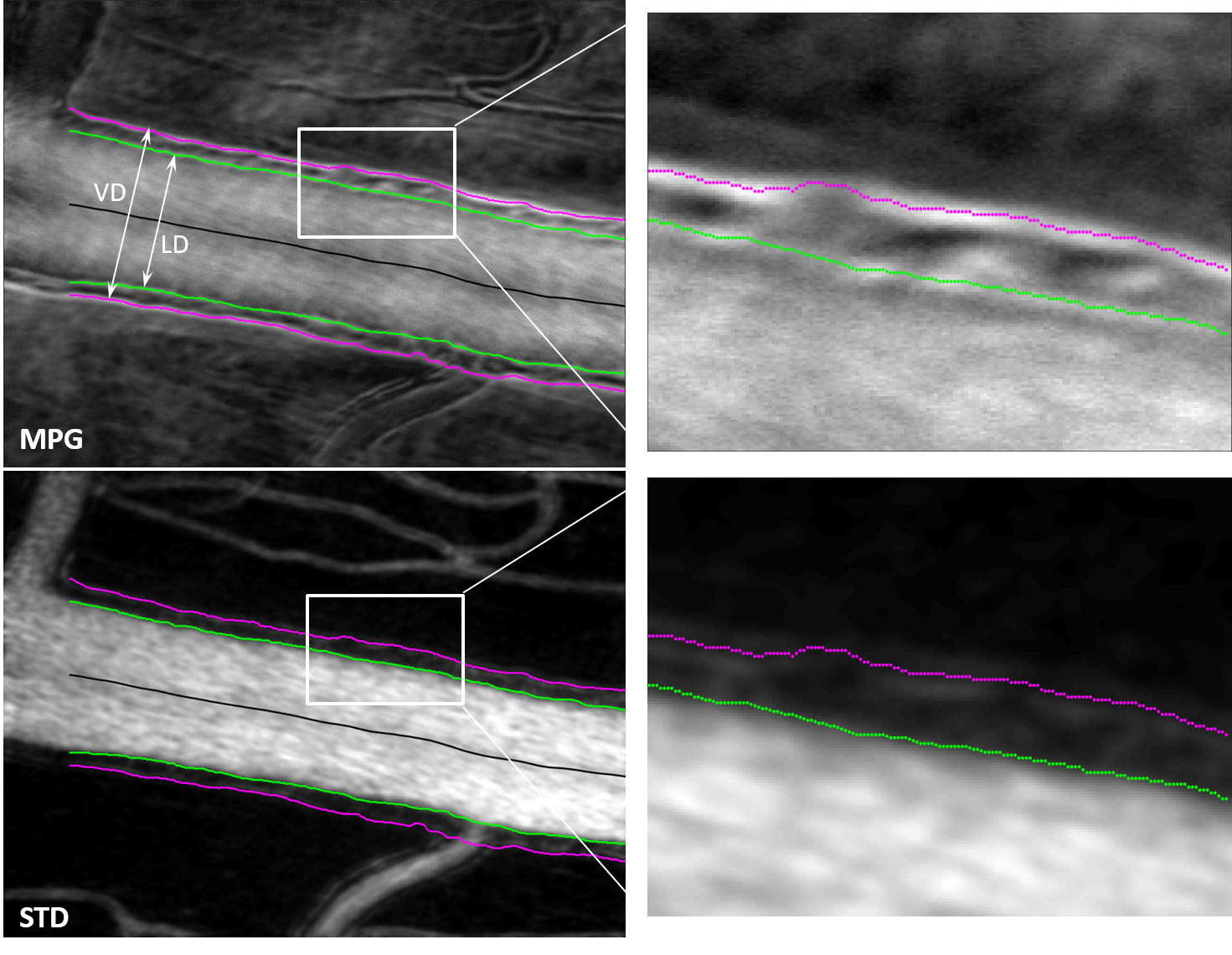
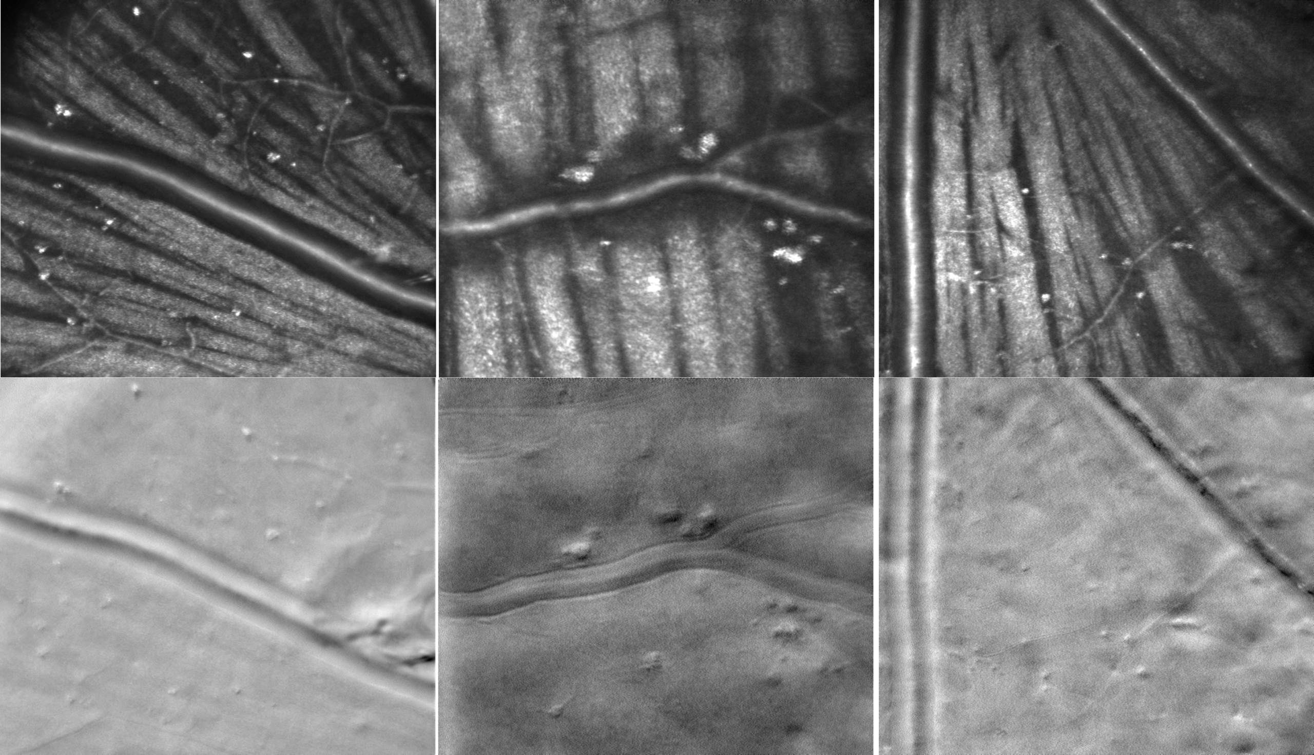
Phase gradient segmentation | “Cellular-Level Analysis of Retinal Blood Vessel Walls Based on Phase Gradient Images”, Diagnostics, 13(22), 3399, (2023) | |
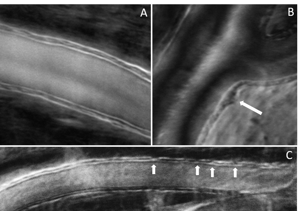
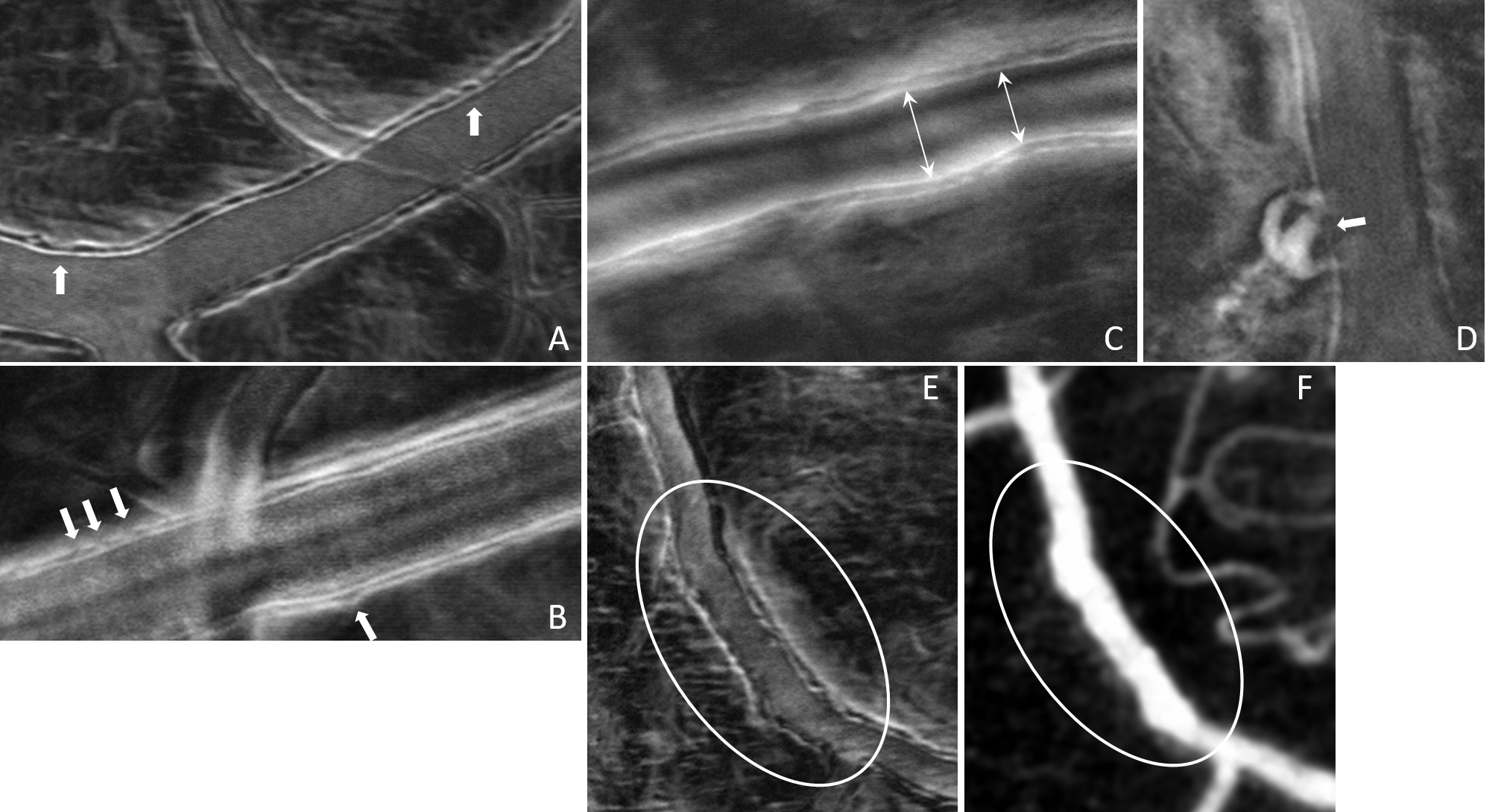
Vessel wall disruptions visualized with phase gradient imaging | “Cellular-Level Analysis of Retinal Blood Vessel Walls Based on Phase Gradient Images”, Diagnostics, 13(22), 3399, (2023) | |
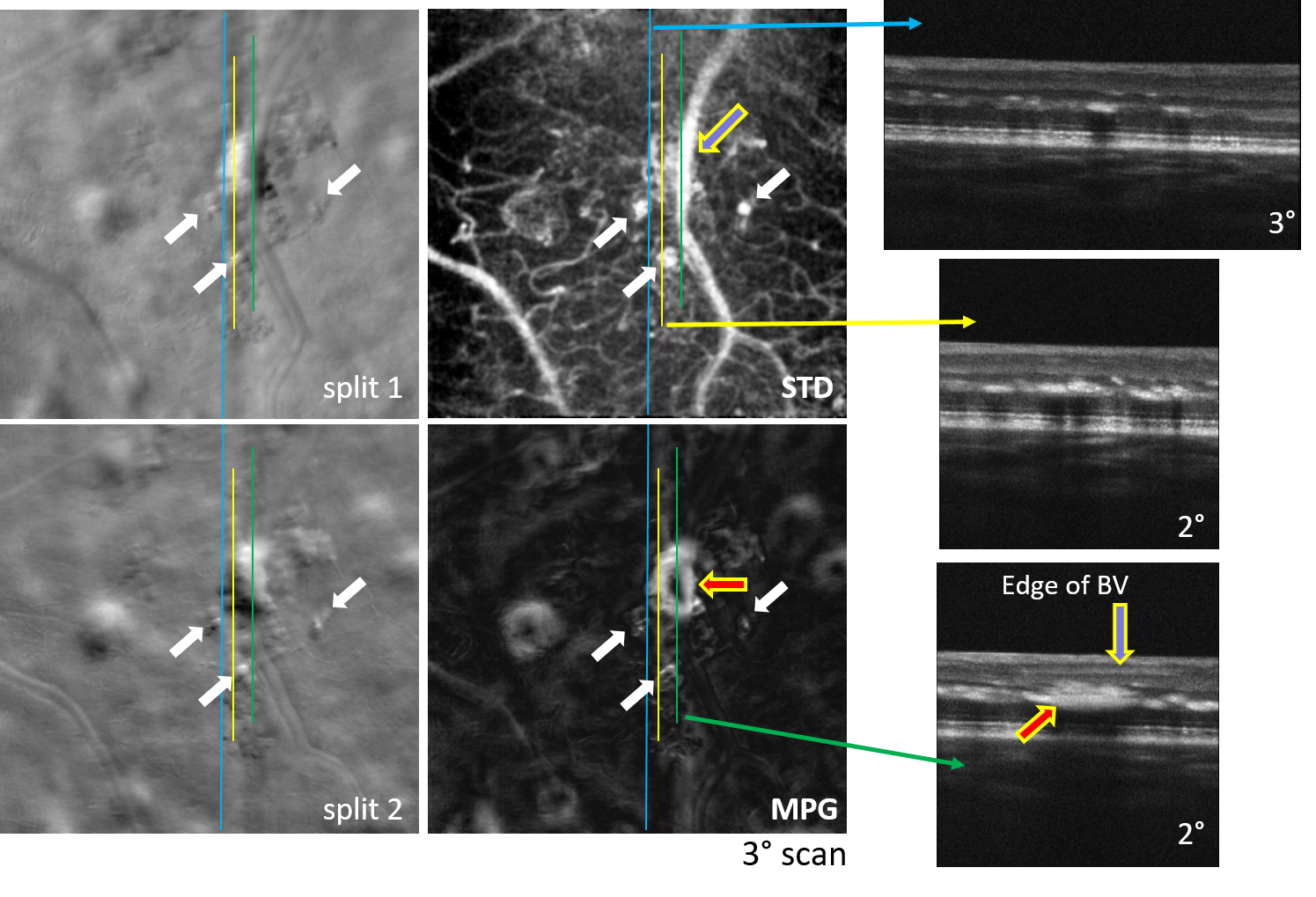
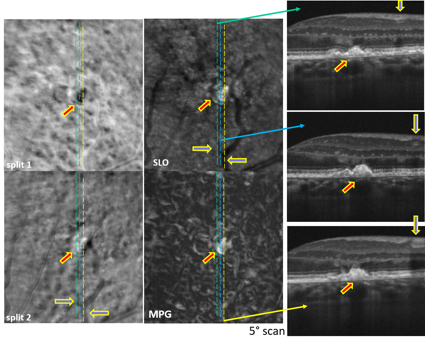
Simultaneous AO-SLO and AO-OCT | Motion Contrast, Phase Gradient, and Simultaneous OCT Images Assist in the Interpretation of Dark-Field Images in Eyes with Retinal Pathology”, Diagnostics, 14(2), 184, (2024) | |
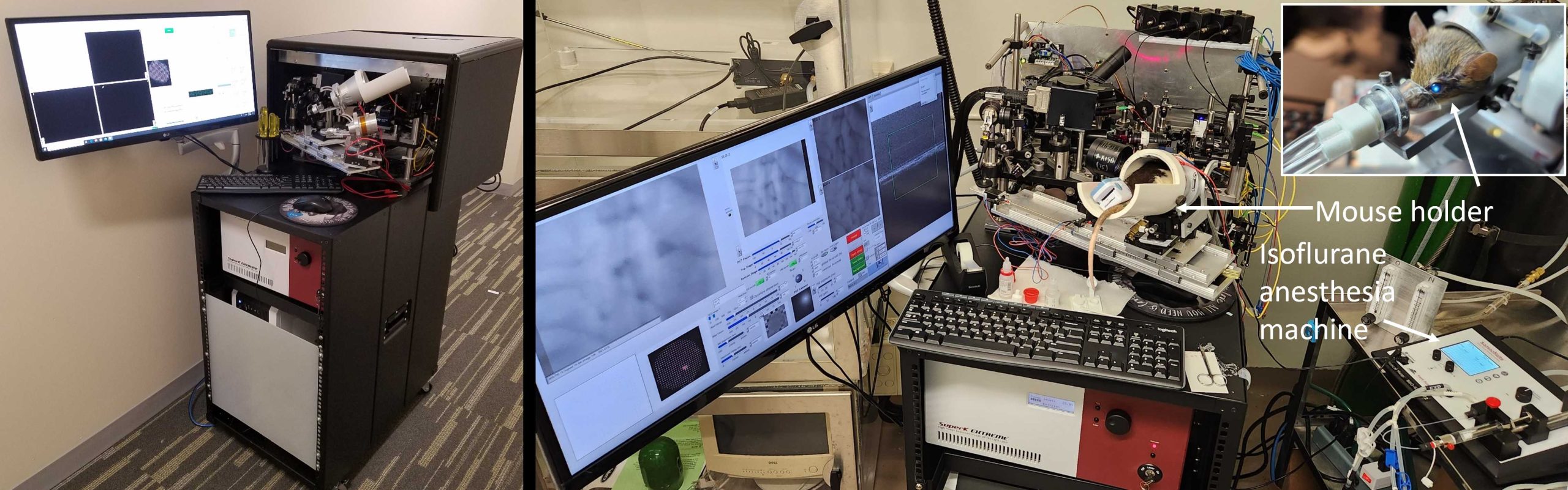
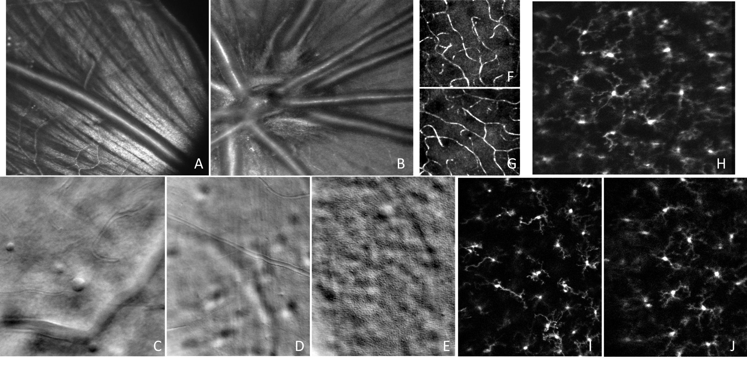
Confocal, offset aperture, motion contrast, and fluorescence small animal retinal imaging | “High-Resolution Retinal Imaging: Technology Overview and Applications”, Photonics 11(6), 522, (2024) | |
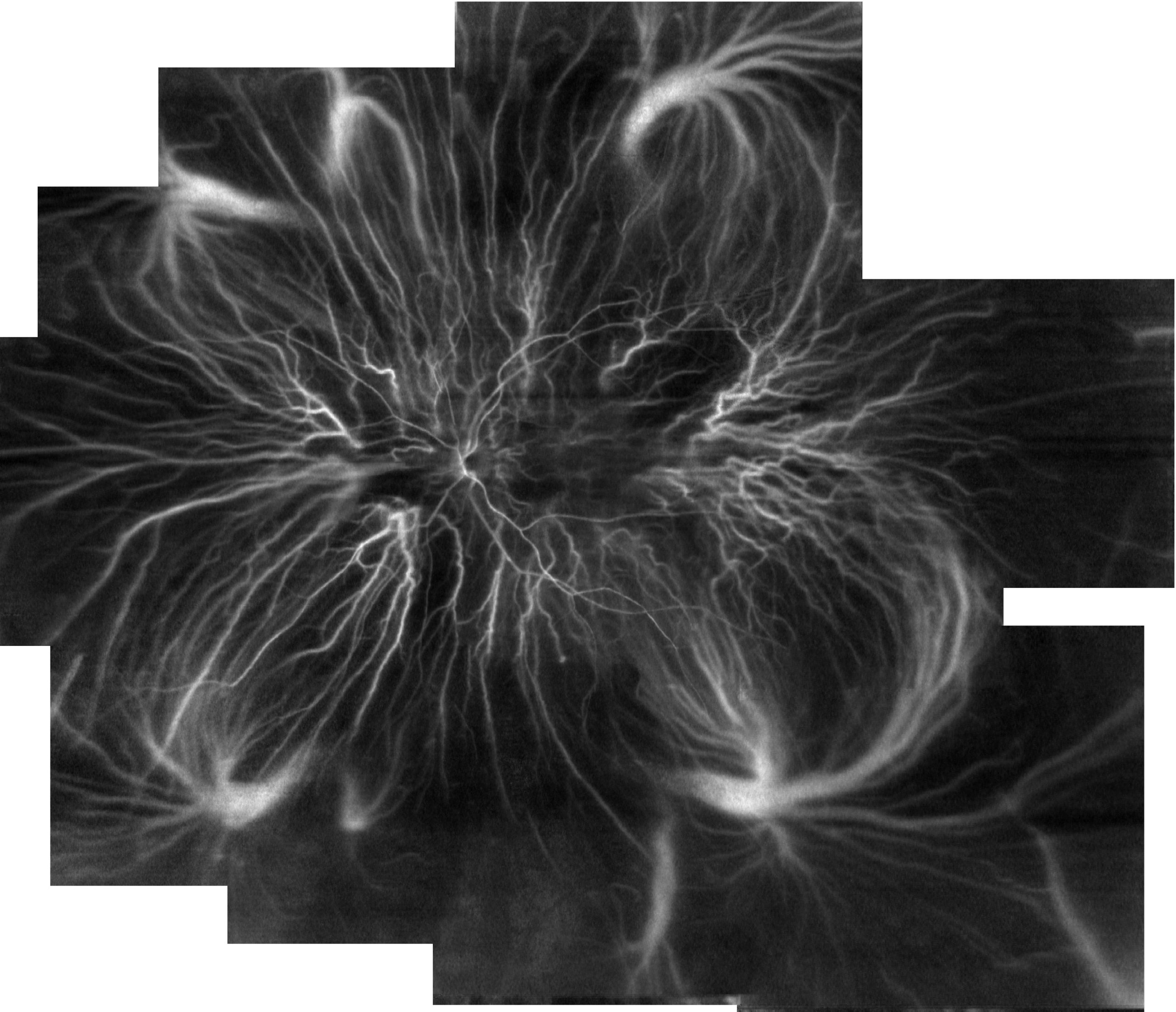
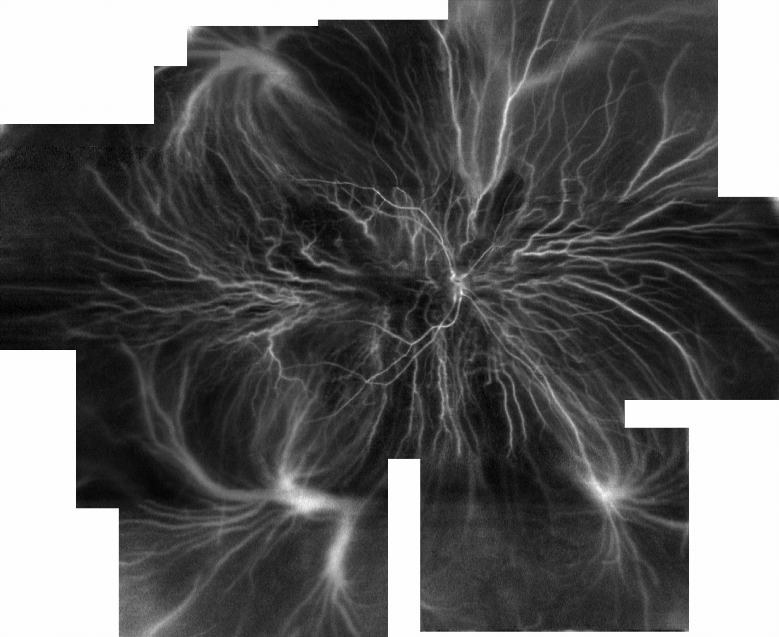
Ultra-wide mapping of retinal and choroidal vasculature with line-scanning Doppler flowmetry | “Visualizing the vasculature of the entire human eye posterior hemisphere without a contrast |